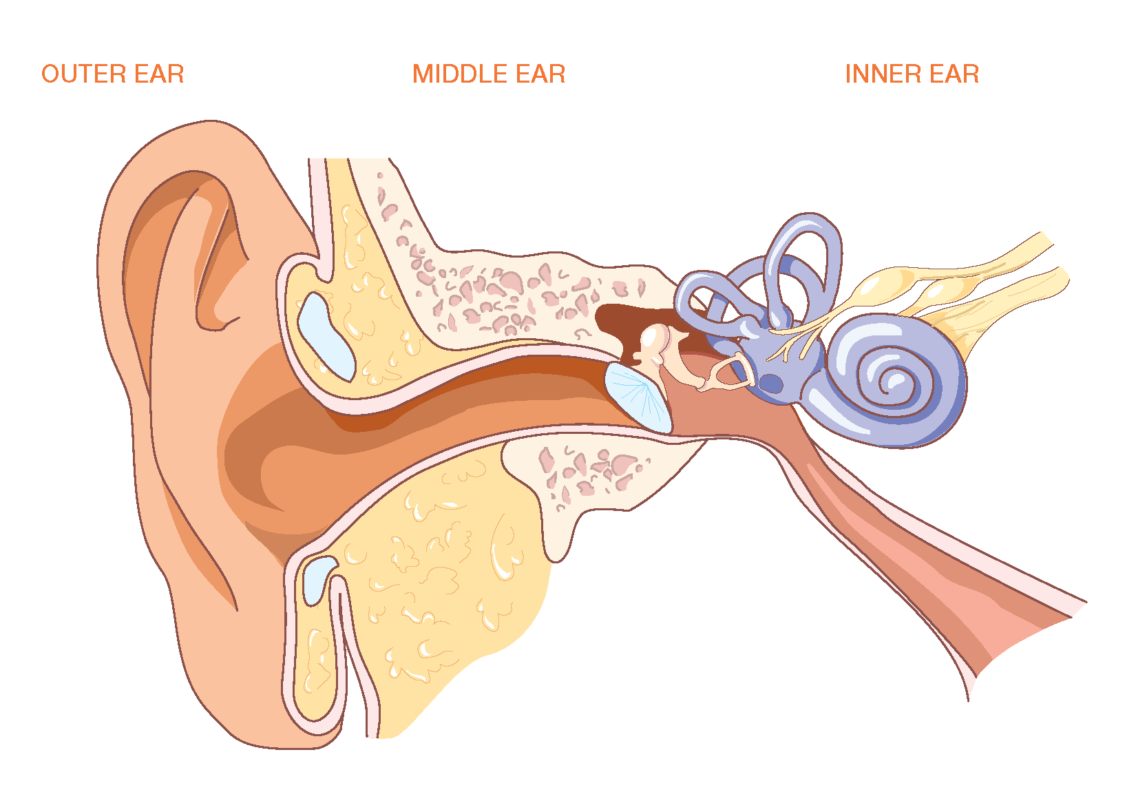
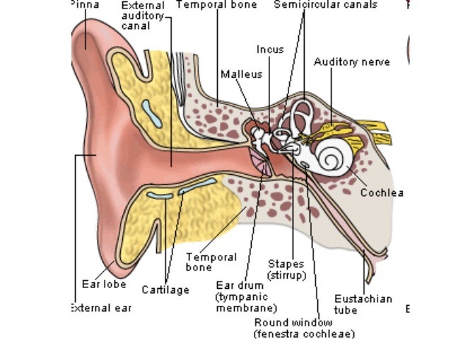
45 picture of human ear with labels
Read About and Diagram the Human Ear | Student Handouts Read About and Diagram the Human Ear Free to Print (PDF) - Science > Upper Elementary Science This free printable worksheet, designed for upper elementary students (grades four, five, and six), has students label a human ear as they read about it. Click here to print. The two pages fit neatly on one double-sided sheet of paper. Human Ear Diagram - Bodytomy The Structure of Human Ear. Helix: It is the prominent outer rim of the external ear. Antihelix: It is the cartilage curve that is situated parallel to the helix. Crus of the Helix: It is the landmark of the outer ear, situated right above the pointy protrusion known as the tragus. Auditory Ossicles: The three small bones in the middle ear ...
Human Ear Photos and Premium High Res Pictures - Getty Images human ear 3d 9,564 Human Ear Premium High Res Photos Browse 9,564 human ear stock photos and images available, or search for human ear close up or human ear anatomy to find more great stock photos and pictures. Related searches: human ear close up human ear anatomy human ear white background human ear diagram human ear icon of 100 NEXT
Picture of human ear with labels
Label of the ear worksheet - Liveworksheets.com Label of the earDrag the parts of the ear to the diagram. ID: 92644. Language: English. School subject: Science. Grade/level: 8. Age: 10-12. Main content: A diagram of the human ear to label. Other contents: Human Ear Anatomy - Parts of Ear Structure, Diagram and Ear Problems The external (outer) ear consists of the auricle, external auditory canal, and eardrum (Figure 1 and 2). The auricle or pinna is a flap of elastic cartilage shaped like the flared end of a trumpet and covered by skin. The rim of the auricle is the helix; the inferior portion is the lobule. Ligaments and muscles attach the auricle to the head. Well-Labelled Diagram Of Ear With Explanation - BYJUS Diagram of Ear. Human ear is a sense organ responsible for hearing and body balance. The outer ear receives the sound waves and transmits them down the ear canal to the eardrum. This causes the eardrum to vibrate and sound is produced. The diagram of ear is important from Class 10 and 12 perspective and is usually asked in the examinations.
Picture of human ear with labels. Human Ear Anatomy Images, Stock Photos & Vectors | Shutterstock human ear anatomy images 12,160 human ear anatomy stock photos, vectors, and illustrations are available royalty-free. See human ear anatomy stock video clips of 122 the earanatomy of earear anatomythe human earanatomy of the earear diagramear structurediagram of earinner middle earhuman ear parts Try these curated collections Next of 122 Human ear diagram, Ear anatomy, Ear diagram - Pinterest See 12 Best Images of Anatomy Human Ear Diagram Worksheet. Inspiring Anatomy Human Ear Diagram Worksheet worksheet images. Worksheeto | Worksheet For You! 434 followers More information Blank Ear Diagram Find this Pin and more on School by Holly Crimm. Ear Anatomy Human Body Anatomy Human Anatomy And Physiology Anatomy Drawing Human Body Organs human body anatomy with labels Stock Photos and Images human body anatomy with labels Stock Photos and Images 4,689 matches Page of 47 The human digestive system, digestive tract or alimentary canal with labels. Labelled with UK spellings and labels like those in the GCSE syllabus Human digestive system, digestive tract or alimentary canal including text labels The Human Ear - Structure, Functions and its Parts - BYJUS There are three ear ossicles in the human ear: Malleus: A hammer-shaped part that is attached to the tympanic membrane through the handle and incus through the head. It is the largest ear ossicle. Incus: An anvil-shaped ear ossicle connected with the stapes. Stapes: It is the smallest ossicle and also the smallest bone in the human body.
Ear Diagram and Labeling Worksheet - Twinkl The first worksheet presents an ear with annotations showing the first letters of its key features. For example, a label marked 'P' links to the Pinna (outer ear). The second page shows an ear diagram without labels. The final page shows the labels linking to the beginning letters of each feature, but without the words list. 244 Human Ear Diagram Premium High Res Photos - Getty Images 244 Human Ear Diagram Premium High Res Photos Browse 244 human ear diagram stock photos and images available, or start a new search to explore more stock photos and images. of 5 NEXT Human Ear: Structure and Functions (With Diagram) ADVERTISEMENTS: In this article we will discuss about the structure and functions of human ear. Structure of Ear: Each ear consists of three portions: (i) External ear, ADVERTISEMENTS: (ii) Middle ear and (iii) Internal ear. 1. External Ear: It comprises a pinna, external auditory meatus (canal) & tympanic membrane. (i) Pinna: ADVERTISEMENTS: The pinna is […] Ear Canal Diagram, Pictures & Anatomy | Body Maps The ear canal, also called the external acoustic meatus, is a passage comprised of bone and skin leading to the eardrum. ... The human ear consists of three regions called the outer ear, middle ...
External Auditory Canal Of Human Ear (With Labels) - Canvas Print Canvas Print Description External Auditory Canal Of Human Ear (With Labels) by Alan Gesek canvas art print arrives ready to hang, with hanging accessories included and no additional framing required. Every canvas print is hand-crafted in the USA, made on-demand at iCanvas and expertly stretched around 100% North American Pine wood stretcher bars. Picture of the Ear: Ear Conditions and Treatments - WebMD Earache: Pain in the ear can have many causes. Some of these are serious, some are not serious. Otitis media (middle ear inflammation): Inflammation or infection of the middle ear (behind the ... Picture Illustration of Ear Anatomy - Ear Structure and Function Picture of Ear The ears and the auditory cortex of the brain are used to perceive sound. The ear is composed of the outer ear, middle ear, and inner ear. Each section performs distinct functions that help transform vibrations into sound. The outer ear is made of skin, cartilage, and bone. It is also the site of the opening to the ear canal. Human Ear Stock Photos, Pictures & Royalty-Free Images - iStock Browse 39,590 human ear stock photos and images available, or search for human ear close up or human ear anatomy to find more great stock photos and pictures. Newest results human ear close up human ear anatomy human ear white background human ear diagram human ear icon human ear illustration human ear line art human ear watercolor human ear sketch
Simple ear diagrams | Ear diagram with labels | Inner ear diagram We provide you here Simple ear diagram in easy way for drawing.Also provided ear diagrams with label and inner ear diagram for better understanding. Also labeled ear diagram available i.e human ear diagram with labels. Diagram of human ear for small kids also provided below. Find this Pin and more on Ear Diagram by ArkLabz. Inner Ear Diagram
Ear Anatomy: Understanding the Outer, Middle, and Inner Parts of the Ear The external auditory meatus, or ear canal, is a narrow canal that leads from the concha to the tympanic membrane, or eardrum. Sound waves are delivered through this canal. This canal is prone to ear infections. Tragus This is the small, rigid part of the ears along the front of the ear, adjacent to the face.
Label the ear | Ear anatomy, Anatomy and physiology, Biology corner The anatomical position. Planes of the human body. Directional terms. The abdominopelvic cavity ear labeling practice The Human Ear - PurposeGames.com - Create. Internal Anatomy of the Ear (58.0K) Anatomy of the Inner Ear (a) (48.0K) Anatomy of the Inner Ear (b) (50.0K)…
External auditory canal of human ear (with labels Stock Photo - Alamy Download this stock image: External auditory canal of human ear (with labels). - E1JKT4 from Alamy's library of millions of high resolution stock photos, illustrations and vectors.
auricle: ear -- Kids Encyclopedia | Children's Homework Help | Kids Online Dictionary | Britannica
Muscle Anatomy With Labels Pictures, Images and Stock Photos Isolated vector illustration on white. muscle anatomy with labels stock illustrations. Synovial joint chart. Labeled anatomy infographic with two bones, Ligaments and Muscles of the Foot, Planar View of the Sole Labeled Parts on White Background muscle anatomy with labels stock pictures, royalty-free photos & images.
Ear Diagram - Human Body Pictures & Images - Science for Kids Photo description: This excellent ear diagram labels all the important parts of the human ear system. The labeled parts include the pinna, auditory canal, eardrum, stapes, malleus, incus and cochlea.
Label Parts of the Human Ear - University of Dayton Label Parts of the Human Ear. Select One Auditory Canal Cochlea Cochlear Nerve Eustachian Tube Incus Malleus Oval Window Pinna Round Window Semicircular Canals Stapes Tympanic Membrane Vestibular Nerve. Select One Auditory Canal Cochlea Cochlear Nerve Eustachian Tube Incus Malleus Oval Window Pinna Round Window Semicircular Canals Stapes ...
Parts of the Ear Labelled Diagram Activity - Twinkl This fun and engaging Parts of the Ear Labelling Activity helps kids identify the different parts of an ear - ideal if you're teaching your class about the body, hearing and physical health. Learners must fill in the labels on the diagram with the appropriate names for each component. This resource is fully differentiated, meaning it comes in three different difficulty levels. This allows ...
12 Best Images of Anatomy Human Ear Diagram Worksheet / worksheeto.com 12 Images of Anatomy Human Ear Diagram Worksheet. Blank Ear Diagram. Human Eye Diagram Unlabeled. General and Special Senses Worksheet. Male and Female Reproductive System Functions. Skeletal System Coloring Pages. Label the Parts of the Heart Worksheet. Parts of the Human Respiratory System. Parts of the Human Respiratory System.
Ear Anatomy, Diagram & Pictures | Body Maps - Healthline Inner ear: The inner ear, also called the labyrinth, operates the body's sense of balance and contains the hearing organ. A bony casing houses a complex system of membranous cells. The inner ear ...
Well-Labelled Diagram Of Ear With Explanation - BYJUS Diagram of Ear. Human ear is a sense organ responsible for hearing and body balance. The outer ear receives the sound waves and transmits them down the ear canal to the eardrum. This causes the eardrum to vibrate and sound is produced. The diagram of ear is important from Class 10 and 12 perspective and is usually asked in the examinations.
Human Ear Anatomy - Parts of Ear Structure, Diagram and Ear Problems The external (outer) ear consists of the auricle, external auditory canal, and eardrum (Figure 1 and 2). The auricle or pinna is a flap of elastic cartilage shaped like the flared end of a trumpet and covered by skin. The rim of the auricle is the helix; the inferior portion is the lobule. Ligaments and muscles attach the auricle to the head.
Label of the ear worksheet - Liveworksheets.com Label of the earDrag the parts of the ear to the diagram. ID: 92644. Language: English. School subject: Science. Grade/level: 8. Age: 10-12. Main content: A diagram of the human ear to label. Other contents:


Post a Comment for "45 picture of human ear with labels"